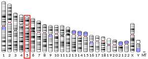
SKP2
SKP2(S-phase kinase associated protein 2)は、ヒトではSKP2遺伝子にコードされるタンパク質である[5][6]。
構造
編集SKP2の全長は424残基であり、N末端領域近傍には約40アミノ酸からなるFボックスドメインが、そしてC末端領域は10個のロイシンリッチリピート(LRR)からなる凹面が形成されている[7]。Fボックスタンパク質は、SCF複合体(SKP1-CUL1-F-box)と呼ばれるユビキチンリガーゼ複合体の4つのサブユニットのうちの1つを構成し、常にではないものの多くの場合、基質をリン酸化依存的に認識する。このSCF複合体において、SKP2は基質認識因子として機能する[8][9][10]。
Fボックスドメイン
編集Fボックスタンパク質は3つのクラスに分類される。FbxwはWD40リピートドメイン、FbxlはLRRをそれぞれ持ち、Fbxoはこれらとは異なる相互作用モジュールを持つか、または識別可能なモチーフを持たないものである[11]。SKP2はFボックスに加えて10個のタンデムなLRRを有し、そのためFbxlに属する。10番目のLRRの後には約30残基のC末端テールが存在し、1番目のLRRへ向かってターンしている。この構造はsafety-beltと呼ばれ、LRRによって形成された凹面へ基質を押しつける役割を果たしている可能性がある[12]。
機能
編集SKP2はサイクリンA-CDK2と安定な複合体を形成する。SKP2は主にS期、G2期、M期の序盤に、リン酸化されたp27(CDKN1B、KIP1)を認識し、分解を促進する[13][14]。SKP2を介したp27の分解は、補助タンパク質CKS1Bを必要とする[15][16]。p27の時期尚早な分解を防ぐため、SKP2の濃度はG1期の序盤から中盤かけて、APC/CCdh1ユビキチンリガーゼによるSKP2のユビキチン化によって低く維持されている[17][18]。SKP2のSer64のリン酸化、そして程度は低いもののSer72のリン酸化はAPC/CCdh1への結合を阻害し、SKP2の安定化に寄与する。一方これらの残基のリン酸化は、SKP2の細胞内局在や活性型SCF複合体への組み立てには必要ではない[19][20][21][22][23]。
細胞周期調節における役割
編集細胞周期の進行は、サイクリン依存性キナーゼ(CDK)、そしてサイクリン、CDK阻害因子との相互作用によって緊密に調節されている。これらによるシグナルの相対量は、周期的なタンパク質分解によって細胞周期の各段階を通じて振動的に増減している[24]。こうした有糸分裂調節タンパク質の分解はユビキチン-プロテアソーム系によって媒介され、細胞内濃度の制御が行われている[25][26]。これらのタンパク質は、E1(ユビキチン活性化酵素)、E2(ユビキチン結合酵素)、E3(ユビキチンリガーゼ)の3つの酵素の逐次的作用によって、認識され分解される[27]。ユビキチン化の特異性をもたらしているのはE3リガーゼであり、E3は標的基質と物理的に相互作用する。SKP2はSCF複合体において基質のリクルートを担う構成要素であり、p27やp21といった細胞周期制御タンパク質を標的としている[28][29][30]。SKP2はp21やp27の双方と二重のネガティブフィードバックループを形成していることが示唆されており、この機構によって細胞周期の開始やG1/S期の移行が制御されている[31][32]。
臨床的意義
編集SKP2はがん遺伝子として振る舞い[33]、リンパ腫の発症に関与するがん原遺伝子としての因果関係が確立されている[34]。がんの発症に関与する最も重要なCDK阻害因子はp27であり、主にサイクリンE-CDK2複合体の阻害(そして程度は低いもののサイクリンD-CDK4複合体の阻害)に関与している。p27の濃度は(他のCDK阻害因子と同様に)細胞周期からの脱出と再進行に応じて増減する。濃度の調節は転写レベルで行われているのではなく、SCF複合体によるp27の認識とプロテアソーム系による分解へのタグ付けによって行われている[24]。細胞がG0期に移行するとSKP2の濃度は低下してp27は増加し、SKP2とp27には見かけ上の逆相関がみられる[17]。SKP2ががんに重要な役割を果たしており、またがんと関連した薬剤抵抗性にも関与していることを強く示唆するエビデンスが蓄積している[35]。
過剰発現
編集SKP2の過剰発現はヒトのがんのプログレッションや転移において高頻度で観察され、SKP2のがん原遺伝子としての役割を示唆するin vitroやin vivoでのエビデンスが得られている[8]。SKP2の過剰発現は、リンパ腫[36]、前立腺がん[37]、メラノーマ[38]、鼻咽頭がん[39][40]、膵がん[41]、乳がん[42][43]でみられる。さらに乳がんでは、SKP2の過剰発現は予後不良と相関している[44][45]。腫瘍異種移植モデルでは、SKP2の過剰発現によって腫瘍成長や腫瘍形成が促進される[46]。SKP2の不活性化は細胞老化またはアポトーシスの開始によってがんの発生を制限するが、この応答はin vivoでの発がん性条件下でのみ観察される[47][48]。この応答はARF/p53非依存的であり、p27依存的に開始される[47][48]。
Skp2ノックアウトマウスモデルを用いて、PTEN、ARF、pRBの不活化やHER2/neuの過剰発現など、さまざまな腫瘍促進条件下におけるがんの発生にSKP2が必要であることが複数のグループによって示されている[49]。また遺伝的アプローチにより、Skp2の枯渇はp53非依存的な細胞老化の誘導やAktを介した好気性解糖の遮断によってがんの発生を阻害することが複数のマウスモデルで実証されている。Skp2の枯渇によってAktの活性化、Glut1の発現、そしてグルコースの取り込みが損なわれ、がんの発生の促進が行われなくなる[50]。
薬剤標的としての可能性
編集SCF複合体の破壊はp27濃度の上昇をもたらし、異常な細胞増殖を阻害すると考えられるため、SKP2は抗がん剤開発の新たな魅力的な標的として多くの関心を集めている。効果的な阻害剤はSKP2と他の因子との相互作用面を標的として開発を行う必要があり、従来的な酵素阻害剤の開発よりもはるかに困難である。SKP2とその基質であるp27との結合部位を標的とした低分子阻害剤が発見されており、これらはSKP2非依存的にp27の蓄積を誘導し、細胞周期の停止を促進する[51]。SKP1/SKP2相互作用面を標的とした阻害剤も発見されており、p27濃度の回復や細胞生存の抑制、p53非依存的な細胞老化の誘導、複数の動物モデルでの強力な抗腫瘍活性、そしてAktを介して解糖系に影響を及ぼすことが発見されている[52]。SKP2はPTENを欠損したがんの治療標的となる可能性がある[47]。
相互作用
編集SKP2は次に挙げる因子と相互作用することが示されている。
出典
編集- ^ a b c GRCh38: Ensembl release 89: ENSG00000145604 - Ensembl, May 2017
- ^ a b c GRCm38: Ensembl release 89: ENSMUSG00000054115 - Ensembl, May 2017
- ^ Human PubMed Reference:
- ^ Mouse PubMed Reference:
- ^ “Chromosomal mapping of the genes for the human CDK2/cyclin A-associated proteins p19 (SKP1A and SKP1B) and p45 (SKP2)”. Cytogenetics and Cell Genetics 73 (1–2): 104–7. (Jul 1996). doi:10.1159/000134318. PMID 8646875.
- ^ “Entrez Gene: SKP2 S-phase kinase-associated protein 2 (p45)”. 2024年6月29日閲覧。
- ^ “SKP1 connects cell cycle regulators to the ubiquitin proteolysis machinery through a novel motif, the F-box”. Cell 86 (2): 263–74. (July 1996). doi:10.1016/S0092-8674(00)80098-7. PMID 8706131.
- ^ a b “Regulation of Skp2 expression and activity and its role in cancer progression”. TheScientificWorldJournal 10: 1001–15. (2010). doi:10.1100/tsw.2010.89. PMC 5763972. PMID 20526532.
- ^ a b “Structure of the Cul1-Rbx1-Skp1-F boxSkp2 SCF ubiquitin ligase complex”. Nature 416 (6882): 703–9. (April 2002). Bibcode: 2002Natur.416..703Z. doi:10.1038/416703a. PMID 11961546.
- ^ “Regulation of the cell cycle by SCF-type ubiquitin ligases”. Seminars in Cell & Developmental Biology 16 (3): 323–33. (June 2005). doi:10.1016/j.semcdb.2005.02.010. PMID 15840441.
- ^ “The F-box protein family”. Genome Biology 1 (5): REVIEWS3002. (2000). doi:10.1186/gb-2000-1-5-reviews3002. PMC 138887. PMID 11178263.
- ^ “The SCF ubiquitin ligase: insights into a molecular machine”. Nature Reviews Molecular Cell Biology 5 (9): 739–51. (September 2004). doi:10.1038/nrm1471. PMID 15340381.
- ^ “SKP2 is required for ubiquitin-mediated degradation of the CDK inhibitor p27”. Nature Cell Biology 1 (4): 193–9. (August 1999). doi:10.1038/12013. PMID 10559916.
- ^ “p27(Kip1) ubiquitination and degradation is regulated by the SCF(Skp2) complex through phosphorylated Thr187 in p27”. Current Biology 9 (12): 661–4. (June 1999). doi:10.1016/S0960-9822(99)80290-5. PMID 10375532.
- ^ a b c “Three different binding sites of Cks1 are required for p27-ubiquitin ligation”. The Journal of Biological Chemistry 277 (44): 42233–40. (November 2002). doi:10.1074/jbc.M205254200. PMID 12140288.
- ^ a b “The cell-cycle regulatory protein Cks1 is required for SCF(Skp2)-mediated ubiquitinylation of p27”. Nature Cell Biology 3 (3): 321–4. (March 2001). doi:10.1038/35060126. PMID 11231585.
- ^ a b “Control of the SCF(Skp2-Cks1) ubiquitin ligase by the APC/C(Cdh1) ubiquitin ligase”. Nature 428 (6979): 190–3. (March 2004). doi:10.1038/nature02330. PMID 15014502.
- ^ “Degradation of the SCF component Skp2 in cell-cycle phase G1 by the anaphase-promoting complex”. Nature 428 (6979): 194–8. (March 2004). Bibcode: 2004Natur.428..194W. doi:10.1038/nature02381. PMID 15014503.
- ^ “Phosphorylation of Skp2 regulated by CDK2 and Cdc14B protects it from degradation by APC(Cdh1) in G1 phase”. The EMBO Journal 27 (4): 679–91. (February 2008). doi:10.1038/emboj.2008.6. PMC 2262036. PMID 18239684.
- ^ “Phosphorylation of Ser72 is dispensable for Skp2 assembly into an active SCF ubiquitin ligase and its subcellular localization”. Cell Cycle 9 (5): 971–4. (March 2010). doi:10.4161/cc.9.5.10914. PMC 3827631. PMID 20160477.
- ^ “Phosphorylation of Ser72 does not regulate the ubiquitin ligase activity and subcellular localization of Skp2”. Cell Cycle 9 (5): 975–9. (March 2010). doi:10.4161/cc.9.5.10915. PMID 20160482.
- ^ “Phosphorylation by Akt1 promotes cytoplasmic localization of Skp2 and impairs APCCdh1-mediated Skp2 destruction”. Nature Cell Biology 11 (4): 397–408. (April 2009). doi:10.1038/ncb1847. PMC 2910589. PMID 19270695.
- ^ “A comparison between Skp2 and FOXO1 for their cytoplasmic localization by Akt1”. Cell Cycle 9 (5): 1021–2. (March 2010). doi:10.4161/cc.9.5.10916. PMC 2990537. PMID 20160512.
- ^ a b “Recycling the cell cycle: cyclins revisited”. Cell 116 (2): 221–34. (January 2004). doi:10.1016/S0092-8674(03)01080-8. PMID 14744433.
- ^ “Themes and variations on ubiquitylation”. Nature Reviews Molecular Cell Biology 2 (3): 169–78. (March 2001). doi:10.1038/35056563. PMID 11265246.
- ^ “Back to the future with ubiquitin”. Cell 116 (2): 181–90. (January 2004). doi:10.1016/S0092-8674(03)01074-2. PMID 14744430.
- ^ “Deregulated proteolysis by the F-box proteins SKP2 and beta-TrCP: tipping the scales of cancer”. Nature Reviews. Cancer 8 (6): 438–49. (June 2008). doi:10.1038/nrc2396. PMC 2711846. PMID 18500245.
- ^ Yu, Z.-K.; Gervais, J. L. M.; Zhang, H. (1998). “Human CUL-1 associates with the SKP1/SKP2 complex and regulates p21CIP1/WAF1 and cyclin D proteins”. Proceedings of the National Academy of Sciences 95 (19): 11324–11329. Bibcode: 1998PNAS...9511324Y. doi:10.1073/pnas.95.19.11324. PMC 21641. PMID 9736735.
- ^ Bornstein, G.; Bloom, J.; Sitry-Shevah, D.; Nakayama, K.; Pagano, M.; Hershko, A. (2003). “Role of the SCFSkp2 Ubiquitin Ligase in the Degradation of p21Cip1 in S Phase”. Journal of Biological Chemistry 278 (28): 25752–25757. doi:10.1074/jbc.m301774200. PMID 12730199.
- ^ Kossatz, U. (2004). “Skp2-dependent degradation of p27kip1 is essential for cell cycle progression”. Genes & Development 18 (21): 2602–2607. doi:10.1101/gad.321004. PMC 525540. PMID 15520280.
- ^ Barr, Alexis R.; Heldt, Frank S.; Zhang, Tongli; Bakal, Chris; Novakl, Bela (2016). “A Dynamical Framework for the All-or-None G1/S Transition”. Cell Systems 2 (1): 27–37. doi:10.1016/j.cels.2016.01.001. PMC 4802413. PMID 27136687.
- ^ Barr, Alexis R.; Cooper, Samuel; Heldt, Frank S.; Butera, Francesca; Stoy, Henriette; Mansfeld, Jorg; Novak, Bela; Bakal, Chris (2017). “DNA damage during S-phase mediates the proliferation-quiescence decision in the subsequent G1 via p21 expression”. Nature Communications 8: 14728. Bibcode: 2017NatCo...814728B. doi:10.1038/ncomms14728. PMC 5364389. PMID 28317845.
- ^ “Role of the F-box protein Skp2 in adhesion-dependent cell cycle progression”. The Journal of Cell Biology 153 (7): 1381–90. (June 2001). doi:10.1083/jcb.153.7.1381. PMC 2150734. PMID 11425869.
- ^ “Role of the F-box protein Skp2 in lymphomagenesis”. Proceedings of the National Academy of Sciences of the United States of America 98 (5): 2515–20. (February 2001). Bibcode: 2001PNAS...98.2515L. doi:10.1073/pnas.041475098. PMC 30169. PMID 11226270.
- ^ “Emerging Roles of SKP2 in Cancer Drug Resistance”. Cells 10 (5): 1147. (May 2021). doi:10.3390/cells10051147. PMC 8150781. PMID 34068643.
- ^ “Prognostic significance of the F-box protein Skp2 expression in diffuse large B-cell lymphoma”. American Journal of Hematology 73 (4): 230–5. (August 2003). doi:10.1002/ajh.10379. PMID 12879424.
- ^ “Skp2: a novel potential therapeutic target for prostate cancer”. Biochimica et Biophysica Acta (BBA) - Reviews on Cancer 1825 (1): 11–7. (January 2012). doi:10.1016/j.bbcan.2011.09.002. PMC 3242930. PMID 21963805.
- ^ “Clinical relevance of SKP2 alterations in metastatic melanoma”. Pigment Cell & Melanoma Research 24 (1): 197–206. (February 2011). doi:10.1111/j.1755-148X.2010.00784.x. PMC 3341662. PMID 20883453.
- ^ “Effect of S-phase kinase-associated protein 2 expression on distant metastasis and survival in nasopharyngeal carcinoma patients”. International Journal of Radiation Oncology, Biology, Physics 73 (1): 202–7. (January 2009). doi:10.1016/j.ijrobp.2008.04.008. PMID 18538504.
- ^ “Correlation of Skp2 overexpression to prognosis of patients with nasopharyngeal carcinoma from South China”. Chinese Journal of Cancer 30 (3): 204–12. (March 2011). doi:10.5732/cjc.010.10403. PMC 4013317. PMID 21352698.
- ^ “SKP2 confers resistance of pancreatic cancer cells towards TRAIL-induced apoptosis”. International Journal of Oncology 38 (1): 219–25. (January 2011). doi:10.3892/ijo_00000841. PMID 21109943.
- ^ “Differential expression of the F-box proteins Skp2 and Skp2B in breast cancer”. Oncogene 24 (21): 3448–58. (May 2005). doi:10.1038/sj.onc.1208328. PMID 15782142.
- ^ “Relationship between levels of Skp2 and P27 in breast carcinomas and possible role of Skp2 as targeted therapy”. Steroids 70 (11): 770–4. (October 2005). doi:10.1016/j.steroids.2005.04.012. PMID 16024059.
- ^ “Significance of skp2 expression in primary breast cancer”. Clinical Cancer Research 12 (4): 1215–20. (February 2006). doi:10.1158/1078-0432.CCR-05-1709. PMID 16489076.
- ^ “Oncogenic role of the ubiquitin ligase subunit Skp2 in human breast cancer”. The Journal of Clinical Investigation 110 (5): 633–41. (September 2002). doi:10.1172/JCI15795. PMC 151109. PMID 12208864.
- ^ “Phosphorylation-dependent regulation of cytosolic localization and oncogenic function of Skp2 by Akt/PKB”. Nature Cell Biology 11 (4): 420–32. (April 2009). doi:10.1038/ncb1849. PMC 2830812. PMID 19270694.
- ^ a b c “Skp2 targeting suppresses tumorigenesis by Arf-p53-independent cellular senescence”. Nature 464 (7287): 374–9. (March 2010). Bibcode: 2010Natur.464..374L. doi:10.1038/nature08815. PMC 2928066. PMID 20237562.
- ^ a b “Disabling Skp2 gene helps shut down cancer growth” (英語). ScienceDaily. 2024年6月30日閲覧。
- ^ “Tumor suppressor ARF inhibits HER-2/neu-mediated oncogenic growth”. Oncogene 23 (42): 7132–43. (September 2004). doi:10.1038/sj.onc.1207918. PMID 15273726.
- ^ “The Skp2-SCF E3 ligase regulates Akt ubiquitination, glycolysis, herceptin sensitivity, and tumorigenesis”. Cell 149 (5): 1098–111. (May 2012). doi:10.1016/j.cell.2012.02.065. PMC 3586339. PMID 22632973.
- ^ “Specific small molecule inhibitors of Skp2-mediated p27 degradation”. Chemistry & Biology 19 (12): 1515–24. (December 2012). doi:10.1016/j.chembiol.2012.09.015. PMC 3530153. PMID 23261596.
- ^ “Pharmacological inactivation of Skp2 SCF ubiquitin ligase restricts cancer stem cell traits and cancer progression”. Cell 154 (3): 556–68. (August 2013). doi:10.1016/j.cell.2013.06.048. PMC 3845452. PMID 23911321.
- ^ a b “Tuberin binds p27 and negatively regulates its interaction with the SCF component Skp2”. The Journal of Biological Chemistry 279 (47): 48707–15. (November 2004). doi:10.1074/jbc.M405528200. PMID 15355997.
- ^ a b c d “Interaction between ubiquitin-protein ligase SCFSKP2 and E2F-1 underlies the regulation of E2F-1 degradation”. Nature Cell Biology 1 (1): 14–9. (May 1999). doi:10.1038/8984. PMID 10559858.
- ^ “Regulation of cyclin A-Cdk2 by SCF component Skp1 and F-box protein Skp2”. Molecular and Cellular Biology 19 (1): 635–45. (January 1999). doi:10.1128/mcb.19.1.635. PMC 83921. PMID 9858587.
- ^ “Role of the SCFSkp2 ubiquitin ligase in the degradation of p21Cip1 in S phase”. The Journal of Biological Chemistry 278 (28): 25752–7. (July 2003). doi:10.1074/jbc.M301774200. PMID 12730199.
- ^ a b “A negatively charged amino acid in Skp2 is required for Skp2-Cks1 interaction and ubiquitination of p27Kip1”. The Journal of Biological Chemistry 278 (34): 32390–6. (August 2003). doi:10.1074/jbc.M305241200. PMID 12813041.
- ^ “Akt-dependent phosphorylation of p27Kip1 promotes binding to 14-3-3 and cytoplasmic localization”. The Journal of Biological Chemistry 277 (32): 28706–13. (August 2002). doi:10.1074/jbc.M203668200. PMID 12042314.
- ^ “Dual-specificity phosphatase 1 ubiquitination in extracellular signal-regulated kinase-mediated control of growth in human hepatocellular carcinoma”. Cancer Research 68 (11): 4192–200. (June 2008). doi:10.1158/0008-5472.CAN-07-6157. PMID 18519678.
- ^ “Structural basis of the Cks1-dependent recognition of p27(Kip1) by the SCF(Skp2) ubiquitin ligase”. Molecular Cell 20 (1): 9–19. (October 2005). doi:10.1016/j.molcel.2005.09.003. PMID 16209941.
- ^ a b “The SCF(Skp2) ubiquitin ligase complex interacts with the human replication licensing factor Cdt1 and regulates Cdt1 degradation”. The Journal of Biological Chemistry 278 (33): 30854–8. (August 2003). doi:10.1074/jbc.C300251200. PMID 12840033.
- ^ a b “TIP120A associates with cullins and modulates ubiquitin ligase activity”. The Journal of Biological Chemistry 278 (18): 15905–10. (May 2003). doi:10.1074/jbc.M213070200. PMID 12609982.
- ^ “Association of human CUL-1 and ubiquitin-conjugating enzyme CDC34 with the F-box protein p45(SKP2): evidence for evolutionary conservation in the subunit composition of the CDC34-SCF pathway”. The EMBO Journal 17 (2): 368–83. (January 1998). doi:10.1093/emboj/17.2.368. PMC 1170388. PMID 9430629.
- ^ “Association of SAP130/SF3b-3 with Cullin-RING ubiquitin ligase complexes and its regulation by the COP9 signalosome”. BMC Biochemistry 9: 1. (2008). doi:10.1186/1471-2091-9-1. PMC 2265268. PMID 18173839.
- ^ “Human origin recognition complex large subunit is degraded by ubiquitin-mediated proteolysis after initiation of DNA replication”. Molecular Cell 9 (3): 481–91. (March 2002). doi:10.1016/S1097-2765(02)00467-7. PMID 11931757.
- ^ “SCF(beta-TRCP) and phosphorylation dependent ubiquitinationof I kappa B alpha catalyzed by Ubc3 and Ubc4”. Oncogene 19 (31): 3529–36. (July 2000). doi:10.1038/sj.onc.1203647. PMID 10918611.
- ^ “Characterization of the cullin and F-box protein partner Skp1”. FEBS Letters 438 (3): 183–9. (November 1998). doi:10.1016/S0014-5793(98)01299-X. PMID 9827542.
- ^ “Insights into SCF ubiquitin ligases from the structure of the Skp1-Skp2 complex”. Nature 408 (6810): 381–6. (November 2000). Bibcode: 2000Natur.408..381S. doi:10.1038/35042620. PMID 11099048.
- ^ “Identification of a family of human F-box proteins”. Current Biology 9 (20): 1177–9. (October 1999). doi:10.1016/S0960-9822(00)80020-2. PMID 10531035.


