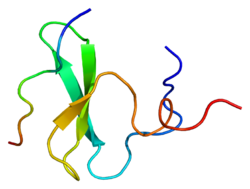
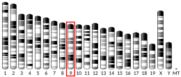
YAP1
YAP1(yes-associated protein 1)は、転写因子として機能するタンパク質であり、細胞増殖に関与する遺伝子の転写を活性化しアポトーシスに関与する遺伝子を抑制する。YAP、YAP65とも呼ばれる。YAP1は、器官のサイズの制御や腫瘍の抑制を可能にするHippoシグナル伝達経路によって阻害される。YAP1はYesやSrcといったチロシンキナーゼのSH3ドメインに結合することから初めて同定された[5]。YAP1をコードするYAP1遺伝子は強力ながん遺伝子であり、ヒトのさまざまながんで増幅されている[6][7]。
構造
編集YAP1遺伝子のクローニングは、WWドメインと呼ばれるモジュール状のタンパク質ドメインの同定を助けた[8][9][10]。YAP1遺伝子産物から同定された2つのスプライシングアイソフォームはYAP1-1、YAP1-2と命名されたが、これらはWWドメインをコードする余分な38アミノ酸が存在するかどうかが異なっていた[11][12]。YAP1の構造は、最もN末端にプロリンリッチ領域、続いてTID(TEAD transcription factor interacting domain)が位置する[13]。次に、YAP1-1アイソフォームでは1つ、YAP1-2アイソフォームでは2つのWWドメインが存在し、SH3-BM(Src Homology 3 binding motif)が続く[5][14]。SH3-BMに続いて、TAD(transcription activation domain)とPDZドメイン結合モチーフ(PDZ-BM)が存在する[15][16]。
機能
編集YAP1は転写のコアクチベーターであり[17]、その増殖促進活性と発がん活性は、細胞増殖を促進しアポトーシスを阻害する遺伝子をアップレギュレーションする、TEADファミリーの転写因子と結合することによって駆動される[13][18]。RUNX[17]、SMAD[19][20]、p73[21]、ErbB4[22][23]、TP53BP[24]、LATS1/2[25]、PTPN14[26]、AMOT[27][28][29][30]、ZO1/2[31]など、TEADファミリー以外の機能的パートナーも同定されている。YAP1とその近縁パラログであるTAZ(WWTR1)はHippoがん抑制経路の主要なエフェクターである[32]。この経路が活性化されると、YAP1とTAZはセリン残基がリン酸化され、14-3-3タンパク質によって細胞質に隔離される[32]。Hippo経路が活性化されていない場合には、YAP1/TAZは細胞核へ移行して遺伝子発現を調節する[32]。
YAP1/TAZは剛性センサーとしても作用することが示されており、Hippoシグナル伝達カスケードとは独立して機械的シグナルの伝達を調節する[33]。
YAP1とTAZは転写コアクチベーターであり、DNA結合ドメインは持っていない。その代わりに核内ではTEAD1-4を介して遺伝子発現を調節する。TEAD1-4は配列特異的転写因子であり、Hippo経路の主要な転写アウトプットを媒介する[34]。YAP1/TAZとTEADの相互作用は、転写リプレッサーとして機能するTEAD/VGLL4の相互作用を競合的に阻害し解離させる[35]。YAP1過剰発現のマウスモデルではTEADの標的遺伝子の発現がアップレギュレーションされ、前駆細胞の増殖の増加と組織の過成長がもたらされる[36]。
調節
編集生化学的調節
編集生化学的レベルでは、YAP1はHippoシグナル伝達経路の一部であり、この経路によって調節される。この経路のキナーゼカスケードはYAP1とTAZの不活性化をもたらす[37]。具体的には、TAOキナーゼがSte20様キナーゼMST1/2の活性化ループ(MST1のThr183、MST2のThr180)をリン酸化する[38][39]。活性化されたMST1/2は、LATS1/2のリクルートとリン酸化を助ける足場タンパク質のSAV1とMOB1A/Bをリン酸化する[40][41]。LATS1/2も2つのグループのMAP4Kによってリン酸化される[42][43]。その後、LATS1/2はYAP1とTAZをリン酸化し、14-3-3タンパク質へ結合させることでYAP1とTAZを細胞質へ隔離する[44]。この経路の活性化の結果は、YAP1/YAZの細胞核への移行の制限である。
機械的シグナルによる調節
編集YAP1は細胞外マトリックス(ECM)の剛性、引っ張り、剪断応力、接着表面などの機械的シグナルによって調節され、その調節は細胞骨格の完全性に依存している[45]。こうした機械的シグナルによって誘導されるYAP1の局在現象は、核の扁平化(nuclear flattening)による核膜孔のサイズの変化、核膜の機械受容イオンチャネル、タンパク質の機械的安定性や他のさまざまな因子による結果であると考えられている[45]。こうした機械的因子は、核の軟化(nuclear softening)とECMの剛性の増大を介して特定種のがん細胞と関係している[46][47][48]。がん細胞でみられる核の軟化は力に応答した核の扁平化を促進し、YAP1の局在を引き起こすと考えられ、発がん性細胞でのYAP1の過剰発現と増殖の促進の説明となる可能性がある[49]。さらに、腫瘍ではインテグリンシグナル伝達の昂進によるECMの剛性の増大が一般的にみられ[48]、細胞と細胞核を扁平化してYAPの核局在の増大を引き起こす可能性がある。反対に、ラミンAの過剰発現など、さまざまな刺激による核の硬化(nuclear stiffening)はYAPの核局在を減少させることが示されている[50][51]。
臨床的意義
編集がんの進行におけるHippoシグナル伝達経路の役割の発見によって、YAP1とTAZには大きな期待と関心が寄せられている[52]。YAP1とTAZの過剰活性化は多くのがんで一般的に観察され、YAP1/TAZを介した転写活性は異常な細胞成長への関与が示唆されている[49][53][54]。しかしながら、YAP1はがん原遺伝子として同定されているものの、細胞の状況に依存してがん抑制遺伝子としても機能することが示されている[55][56]。
YAP1がん遺伝子は新たな抗がん剤開発の標的となっており[57]、YAP1-TEAD複合体の形成やWWドメインの結合機能を防ぐ低分子化合物が同定されている[58][59]。こうした低分子は、YAP1がん遺伝子の増幅や過剰発現がみられるがん患者に対する治療法開発のためのリード化合物となっている。
YAP1遺伝子のヘテロ接合型機能喪失変異が眼に大きな形成異常を抱える2家族に同定されている。難聴、口唇裂、知的障害、腎臓疾患など眼以外の異常がみられる場合もある[60]。
Hippo/YAPシグナル伝達経路は、脳の虚血/再灌流障害後の血液脳関門の破壊を緩和することで神経保護効果を発揮する可能性がある[61]。
出典
編集- ^ a b c GRCh38: Ensembl release 89: ENSG00000137693 - Ensembl, May 2017
- ^ a b c GRCm38: Ensembl release 89: ENSMUSG00000053110 - Ensembl, May 2017
- ^ Human PubMed Reference:
- ^ Mouse PubMed Reference:
- ^ a b Sudol, M. (1994-08-XX). “Yes-associated protein (YAP65) is a proline-rich phosphoprotein that binds to the SH3 domain of the Yes proto-oncogene product”. Oncogene 9 (8): 2145–2152. ISSN 0950-9232. PMID 8035999.
- ^ Huang, Jianbin; Wu, Shian; Barrera, Jose; Matthews, Krista; Pan, Duojia (2005-08-12). “The Hippo signaling pathway coordinately regulates cell proliferation and apoptosis by inactivating Yorkie, the Drosophila Homolog of YAP”. Cell 122 (3): 421–434. doi:10.1016/j.cell.2005.06.007. ISSN 0092-8674. PMID 16096061.
- ^ Overholtzer, Michael; Zhang, Jianmin; Smolen, Gromoslaw A.; Muir, Beth; Li, Wenmei; Sgroi, Dennis C.; Deng, Chu-Xia; Brugge, Joan S. et al. (2006-08-15). “Transforming properties of YAP, a candidate oncogene on the chromosome 11q22 amplicon”. Proceedings of the National Academy of Sciences of the United States of America 103 (33): 12405–12410. doi:10.1073/pnas.0605579103. ISSN 0027-8424. PMC 1533802. PMID 16894141.
- ^ Bork, P.; Sudol, M. (1994-12-XX). “The WW domain: a signalling site in dystrophin?”. Trends in Biochemical Sciences 19 (12): 531–533. doi:10.1016/0968-0004(94)90053-1. ISSN 0968-0004. PMID 7846762.
- ^ André, B.; Springael, J. Y. (1994-12-15). “WWP, a new amino acid motif present in single or multiple copies in various proteins including dystrophin and the SH3-binding Yes-associated protein YAP65”. Biochemical and Biophysical Research Communications 205 (2): 1201–1205. doi:10.1006/bbrc.1994.2793. ISSN 0006-291X. PMID 7802651.
- ^ Hofmann, K.; Bucher, P. (1995-01-23). “The rsp5-domain is shared by proteins of diverse functions”. FEBS letters 358 (2): 153–157. doi:10.1016/0014-5793(94)01415-w. ISSN 0014-5793. PMID 7828727.
- ^ Sudol, M.; Bork, P.; Einbond, A.; Kastury, K.; Druck, T.; Negrini, M.; Huebner, K.; Lehman, D. (1995-06-16). “Characterization of the mammalian YAP (Yes-associated protein) gene and its role in defining a novel protein module, the WW domain”. The Journal of Biological Chemistry 270 (24): 14733–14741. doi:10.1074/jbc.270.24.14733. ISSN 0021-9258. PMID 7782338.
- ^ Gaffney, Christian J.; Oka, Tsutomu; Mazack, Virginia; Hilman, Dror; Gat, Uri; Muramatsu, Tomoki; Inazawa, Johji; Golden, Alicia et al. (2012-11-10). “Identification, basic characterization and evolutionary analysis of differentially spliced mRNA isoforms of human YAP1 gene”. Gene 509 (2): 215–222. doi:10.1016/j.gene.2012.08.025. ISSN 1879-0038. PMC 3455135. PMID 22939869.
- ^ a b Vassilev, A.; Kaneko, K. J.; Shu, H.; Zhao, Y.; DePamphilis, M. L. (2001-05-15). “TEAD/TEF transcription factors utilize the activation domain of YAP65, a Src/Yes-associated protein localized in the cytoplasm”. Genes & Development 15 (10): 1229–1241. doi:10.1101/gad.888601. ISSN 0890-9369. PMC 313800. PMID 11358867.
- ^ Ren, R.; Mayer, B. J.; Cicchetti, P.; Baltimore, D. (1993-02-19). “Identification of a ten-amino acid proline-rich SH3 binding site”. Science (New York, N.Y.) 259 (5098): 1157–1161. doi:10.1126/science.8438166. ISSN 0036-8075. PMID 8438166.
- ^ Wang, S.; Raab, R. W.; Schatz, P. J.; Guggino, W. B.; Li, M. (1998-05-01). “Peptide binding consensus of the NHE-RF-PDZ1 domain matches the C-terminal sequence of cystic fibrosis transmembrane conductance regulator (CFTR)”. FEBS letters 427 (1): 103–108. doi:10.1016/s0014-5793(98)00402-5. ISSN 0014-5793. PMID 9613608.
- ^ Mohler, P. J.; Kreda, S. M.; Boucher, R. C.; Sudol, M.; Stutts, M. J.; Milgram, S. L. (1999-11-15). “Yes-associated protein 65 localizes p62(c-Yes) to the apical compartment of airway epithelia by association with EBP50”. The Journal of Cell Biology 147 (4): 879–890. doi:10.1083/jcb.147.4.879. ISSN 0021-9525. PMC 2156157. PMID 10562288.
- ^ a b Yagi, R.; Chen, L. F.; Shigesada, K.; Murakami, Y.; Ito, Y. (1999-05-04). “A WW domain-containing yes-associated protein (YAP) is a novel transcriptional co-activator”. The EMBO journal 18 (9): 2551–2562. doi:10.1093/emboj/18.9.2551. ISSN 0261-4189. PMC 1171336. PMID 10228168.
- ^ Zhao, Bin; Kim, Joungmok; Ye, Xin; Lai, Zhi-Chun; Guan, Kun-Liang (2009-02-01). “Both TEAD-binding and WW domains are required for the growth stimulation and oncogenic transformation activity of yes-associated protein”. Cancer Research 69 (3): 1089–1098. doi:10.1158/0008-5472.CAN-08-2997. ISSN 1538-7445. PMID 19141641.
- ^ Ferrigno, Olivier; Lallemand, François; Verrecchia, Franck; L'Hoste, Sébastien; Camonis, Jacques; Atfi, Azeddine; Mauviel, Alain (2002-07-25). “Yes-associated protein (YAP65) interacts with Smad7 and potentiates its inhibitory activity against TGF-beta/Smad signaling”. Oncogene 21 (32): 4879–4884. doi:10.1038/sj.onc.1205623. ISSN 0950-9232. PMID 12118366.
- ^ Aragón, Eric; Goerner, Nina; Xi, Qiaoran; Gomes, Tiago; Gao, Sheng; Massagué, Joan; Macias, Maria J. (2012-10-10). “Structural basis for the versatile interactions of Smad7 with regulator WW domains in TGF-β Pathways”. Structure (London, England: 1993) 20 (10): 1726–1736. doi:10.1016/j.str.2012.07.014. ISSN 1878-4186. PMC 3472128. PMID 22921829.
- ^ Strano, S.; Munarriz, E.; Rossi, M.; Castagnoli, L.; Shaul, Y.; Sacchi, A.; Oren, M.; Sudol, M. et al. (2001-05-04). “Physical interaction with Yes-associated protein enhances p73 transcriptional activity”. The Journal of Biological Chemistry 276 (18): 15164–15173. doi:10.1074/jbc.M010484200. ISSN 0021-9258. PMID 11278685.
- ^ Komuro, Akihiko; Nagai, Makoto; Navin, Nicholas E.; Sudol, Marius (2003-08-29). “WW domain-containing protein YAP associates with ErbB-4 and acts as a co-transcriptional activator for the carboxyl-terminal fragment of ErbB-4 that translocates to the nucleus”. The Journal of Biological Chemistry 278 (35): 33334–33341. doi:10.1074/jbc.M305597200. ISSN 0021-9258. PMID 12807903.
- ^ Omerovic, Jasminka; Puggioni, Eleonora M. R.; Napoletano, Silvia; Visco, Vincenzo; Fraioli, Rocco; Frati, Luigi; Gulino, Alberto; Alimandi, Maurizio (2004-04-01). “Ligand-regulated association of ErbB-4 to the transcriptional co-activator YAP65 controls transcription at the nuclear level”. Experimental Cell Research 294 (2): 469–479. doi:10.1016/j.yexcr.2003.12.002. ISSN 0014-4827. PMID 15023535.
- ^ Espanel, X.; Sudol, M. (2001-04-27). “Yes-associated protein and p53-binding protein-2 interact through their WW and SH3 domains”. The Journal of Biological Chemistry 276 (17): 14514–14523. doi:10.1074/jbc.M008568200. ISSN 0021-9258. PMID 11278422.
- ^ Oka, Tsutomu; Mazack, Virginia; Sudol, Marius (2008-10-10). “Mst2 and Lats kinases regulate apoptotic function of Yes kinase-associated protein (YAP)”. The Journal of Biological Chemistry 283 (41): 27534–27546. doi:10.1074/jbc.M804380200. ISSN 0021-9258. PMID 18640976.
- ^ Liu, X.; Yang, N.; Figel, S. A.; Wilson, K. E.; Morrison, C. D.; Gelman, I. H.; Zhang, J. (2013-03-07). “PTPN14 interacts with and negatively regulates the oncogenic function of YAP”. Oncogene 32 (10): 1266–1273. doi:10.1038/onc.2012.147. ISSN 1476-5594. PMC 4402938. PMID 22525271.
- ^ Wang, Wenqi; Huang, Jun; Chen, Junjie (2011-02-11). “Angiomotin-like proteins associate with and negatively regulate YAP1”. The Journal of Biological Chemistry 286 (6): 4364–4370. doi:10.1074/jbc.C110.205401. ISSN 1083-351X. PMC 3039387. PMID 21187284.
- ^ Chan, Siew Wee; Lim, Chun Jye; Chong, Yaan Fun; Pobbati, Ajaybabu V.; Huang, Caixia; Hong, Wanjin (2011-03-04). “Hippo pathway-independent restriction of TAZ and YAP by angiomotin”. The Journal of Biological Chemistry 286 (9): 7018–7026. doi:10.1074/jbc.C110.212621. ISSN 1083-351X. PMC 3044958. PMID 21224387.
- ^ Zhao, Bin; Li, Li; Lu, Qing; Wang, Lloyd H.; Liu, Chen-Ying; Lei, Qunying; Guan, Kun-Liang (2011-01-01). “Angiomotin is a novel Hippo pathway component that inhibits YAP oncoprotein”. Genes & Development 25 (1): 51–63. doi:10.1101/gad.2000111. ISSN 1549-5477. PMC 3012936. PMID 21205866.
- ^ Oka, T.; Schmitt, A. P.; Sudol, M. (2012-01-05). “Opposing roles of angiomotin-like-1 and zona occludens-2 on pro-apoptotic function of YAP”. Oncogene 31 (1): 128–134. doi:10.1038/onc.2011.216. ISSN 1476-5594. PMID 21685940.
- ^ Oka, Tsutomu; Remue, Eline; Meerschaert, Kris; Vanloo, Berlinda; Boucherie, Ciska; Gfeller, David; Bader, Gary D.; Sidhu, Sachdev S. et al. (2010-12-15). “Functional complexes between YAP2 and ZO-2 are PDZ domain-dependent, and regulate YAP2 nuclear localization and signalling”. The Biochemical Journal 432 (3): 461–472. doi:10.1042/BJ20100870. ISSN 1470-8728. PMID 20868367.
- ^ a b c Pan, Duojia (2010-10-19). “The hippo signaling pathway in development and cancer”. Developmental Cell 19 (4): 491–505. doi:10.1016/j.devcel.2010.09.011. ISSN 1878-1551. PMC 3124840. PMID 20951342.
- ^ “Using biomaterials to study stem cell mechanotransduction, growth and differentiation”. Journal of Tissue Engineering and Regenerative Medicine 9 (5): 528–39. (May 2015). doi:10.1002/term.1957. PMID 25370612.
- ^ “TEAD mediates YAP-dependent gene induction and growth control”. Genes & Development 22 (14): 1962–71. (July 2008). doi:10.1101/gad.1664408. PMC 2492741. PMID 18579750.
- ^ “The Hippo effector Yorkie controls normal tissue growth by antagonizing scalloped-mediated default repression”. Developmental Cell 25 (4): 388–401. (May 2013). doi:10.1016/j.devcel.2013.04.021. PMC 3705890. PMID 23725764.
- ^ “Homeostatic control of Hippo signaling activity revealed by an endogenous activating mutation in YAP”. Genes & Development 29 (12): 1285–97. (June 2015). doi:10.1101/gad.264234.115. PMC 4495399. PMID 26109051.
- ^ “Mechanisms of Hippo pathway regulation”. Genes & Development 30 (1): 1–17. (January 2016). doi:10.1101/gad.274027.115. PMC 4701972. PMID 26728553.
- ^ “Tao-1 phosphorylates Hippo/MST kinases to regulate the Hippo-Salvador-Warts tumor suppressor pathway”. Developmental Cell 21 (5): 888–95. (November 2011). doi:10.1016/j.devcel.2011.08.028. PMC 3217187. PMID 22075147.
- ^ “The sterile 20-like kinase Tao-1 controls tissue growth by regulating the Salvador-Warts-Hippo pathway”. Developmental Cell 21 (5): 896–906. (November 2011). doi:10.1016/j.devcel.2011.09.012. PMID 22075148.
- ^ “Association of mammalian sterile twenty kinases, Mst1 and Mst2, with hSalvador via C-terminal coiled-coil domains, leads to its stabilization and phosphorylation”. The FEBS Journal 273 (18): 4264–76. (September 2006). doi:10.1111/j.1742-4658.2006.05427.x. PMID 16930133.
- ^ “MOBKL1A/MOBKL1B phosphorylation by MST1 and MST2 inhibits cell proliferation”. Current Biology 18 (5): 311–21. (March 2008). doi:10.1016/j.cub.2008.02.006. PMC 4682548. PMID 18328708.
- ^ “MAP4K family kinases act in parallel to MST1/2 to activate LATS1/2 in the Hippo pathway”. Nature Communications 6: 8357. (October 2015). Bibcode: 2015NatCo...6.8357M. doi:10.1038/ncomms9357. PMC 4600732. PMID 26437443.
- ^ “Identification of Happyhour/MAP4K as Alternative Hpo/Mst-like Kinases in the Hippo Kinase Cascade”. Developmental Cell 34 (6): 642–55. (September 2015). doi:10.1016/j.devcel.2015.08.014. PMC 4589524. PMID 26364751.
- ^ “Inactivation of YAP oncoprotein by the Hippo pathway is involved in cell contact inhibition and tissue growth control”. Genes & Development 21 (21): 2747–61. (November 2007). doi:10.1101/gad.1602907. PMC 2045129. PMID 17974916.
- ^ a b “Force Triggers YAP Nuclear Entry by Regulating Transport across Nuclear Pores”. Cell 171 (6): 1397–1410.e14. (November 2017). doi:10.1016/j.cell.2017.10.008. PMID 29107331.
- ^ “Nanomechanical analysis of cells from cancer patients”. Nature Nanotechnology 2 (12): 780–3. (December 2007). Bibcode: 2007NatNa...2..780C. doi:10.1038/nnano.2007.388. PMID 18654431.
- ^ “Optical deformability as an inherent cell marker for testing malignant transformation and metastatic competence” (English). Biophysical Journal 88 (5): 3689–98. (May 2005). Bibcode: 2005BpJ....88.3689G. doi:10.1529/biophysj.104.045476. PMC 1305515. PMID 15722433.
- ^ a b “Cancer invasion and the microenvironment: plasticity and reciprocity”. Cell 147 (5): 992–1009. (November 2011). doi:10.1016/j.cell.2011.11.016. PMID 22118458.
- ^ a b “The PDZ-binding motif of Yes-associated protein is required for its co-activation of TEAD-mediated CTGF transcription and oncogenic cell transforming activity”. Biochemical and Biophysical Research Communications 443 (3): 917–23. (January 2014). doi:10.1016/j.bbrc.2013.12.100. PMID 24380865.
- ^ “Nuclear lamin-A scales with tissue stiffness and enhances matrix-directed differentiation”. Science 341 (6149): 1240104. (August 2013). doi:10.1126/science.1240104. PMC 3976548. PMID 23990565.
- ^ “Designer matrices for intestinal stem cell and organoid culture”. Nature 539 (7630): 560–564. (November 2016). doi:10.1038/nature20168. PMID 27851739.
- ^ “The emerging roles of YAP and TAZ in cancer”. Nature Reviews. Cancer 15 (2): 73–79. (February 2015). doi:10.1038/nrc3876. PMC 4562315. PMID 25592648.
- ^ “The Hippo pathway and human cancer”. Nature Reviews. Cancer 13 (4): 246–57. (April 2013). doi:10.1038/nrc3458. PMID 23467301.
- ^ “The two faces of Hippo: targeting the Hippo pathway for regenerative medicine and cancer treatment”. Nature Reviews. Drug Discovery 13 (1): 63–79. (January 2014). doi:10.1038/nrd4161. PMC 4167640. PMID 24336504.
- ^ “Restriction of intestinal stem cell expansion and the regenerative response by YAP”. Nature 493 (7430): 106–10. (January 2013). Bibcode: 2013Natur.493..106B. doi:10.1038/nature11693. PMC 3536889. PMID 23178811.
- ^ “Rescue of Hippo coactivator YAP1 triggers DNA damage-induced apoptosis in hematological cancers”. Nature Medicine 20 (6): 599–606. (June 2014). doi:10.1038/nm.3562. PMC 4057660. PMID 24813251.
- ^ Sudol, Marius; Shields, Denis C.; Farooq, Amjad (2012-09). “Structures of YAP protein domains reveal promising targets for development of new cancer drugs”. Seminars in Cell & Developmental Biology 23 (7): 827–833. doi:10.1016/j.semcdb.2012.05.002. ISSN 1096-3634. PMC 3427467. PMID 22609812.
- ^ Liu-Chittenden, Yi; Huang, Bo; Shim, Joong Sup; Chen, Qian; Lee, Se-Jin; Anders, Robert A.; Liu, Jun O.; Pan, Duojia (2012-06-15). “Genetic and pharmacological disruption of the TEAD-YAP complex suppresses the oncogenic activity of YAP”. Genes & Development 26 (12): 1300–1305. doi:10.1101/gad.192856.112. ISSN 1549-5477. PMC 3387657. PMID 22677547.
- ^ Kang, Seung-gu; Huynh, Tien; Zhou, Ruhong (2012). “Non-destructive inhibition of metallofullerenol Gd@C(82)(OH)(22) on WW domain: implication on signal transduction pathway”. Scientific Reports 2: 957. doi:10.1038/srep00957. ISSN 2045-2322. PMC 3518810. PMID 23233876.
- ^ “Heterozygous loss-of-function mutations in YAP1 cause both isolated and syndromic optic fissure closure defects”. American Journal of Human Genetics 94 (2): 295–302. (February 2014). doi:10.1016/j.ajhg.2014.01.001. PMC 3928658. PMID 24462371.
- ^ “Hippo/YAP signaling pathway mitigates blood-brain barrier disruption after cerebral ischemia/reperfusion injury”. Behavioural Brain Research 356: 8–17. (January 2019). doi:10.1016/j.bbr.2018.08.003. PMC 6193462. PMID 30092249.


